

Authors: Dr Jonathan Dimitry, Dr Housam Ghazzawi. United Lincolnshire Teaching Hospitals NHS Trust
Declaration: This case presentation prepared by us is based on our own clinical work and review of the literature and has not been copied from another source. All patient-identifiable details have been removed from the text and images to protect confidentiality in line with UK GDPR requirements. The patient gave informed consent for his case and relevant imaging to be used for academic/presentation purposes.
Introduction:
Hip fractures are a common reason for hospital admission in older adults and are associated with substantial morbidity, loss of independence, and increased mortality [1,2]. Operative treatment is the mainstay of management for most hip fractures, with the aim of enabling early mobilisation and reducing complications related to prolonged immobility [1].
Myocardial infarction (MI) is a recognised complication after hip fracture surgery and contributes to worse short-term outcomes in this high-risk population [3,4]. Type 2 myocardial infarction (T2MI) is defined as myocardial necrosis caused by an imbalance between myocardial oxygen supply and demand, rather than acute coronary plaque rupture, and common triggers include anaemia, hypoxia, sepsis, hypotension, and other physiological stressors [5,6].
We present a case of an elderly patient who developed a T2MI following intramedullary nailing for a neck of femur fracture, highlighting the challenges of diagnosis and management in the immediate post-operative setting.
Article:
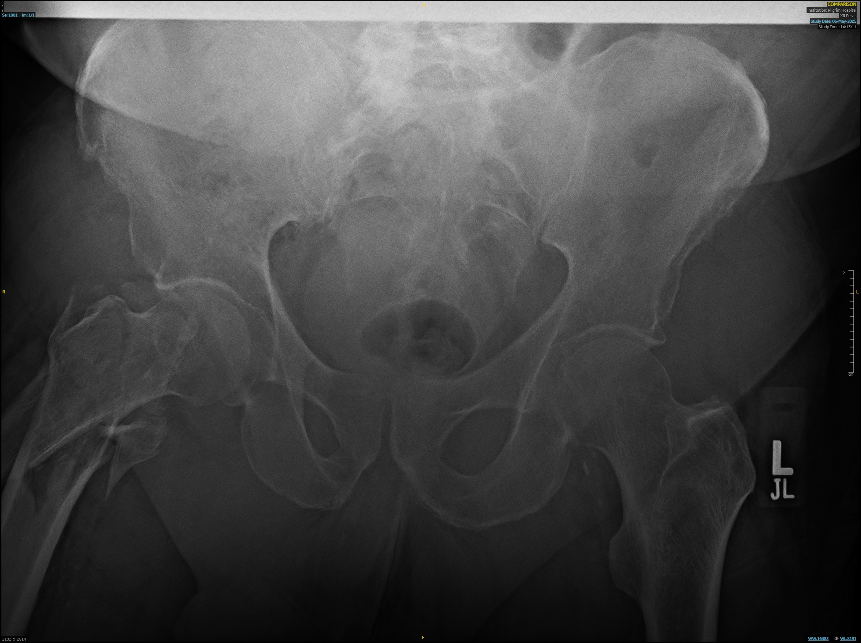
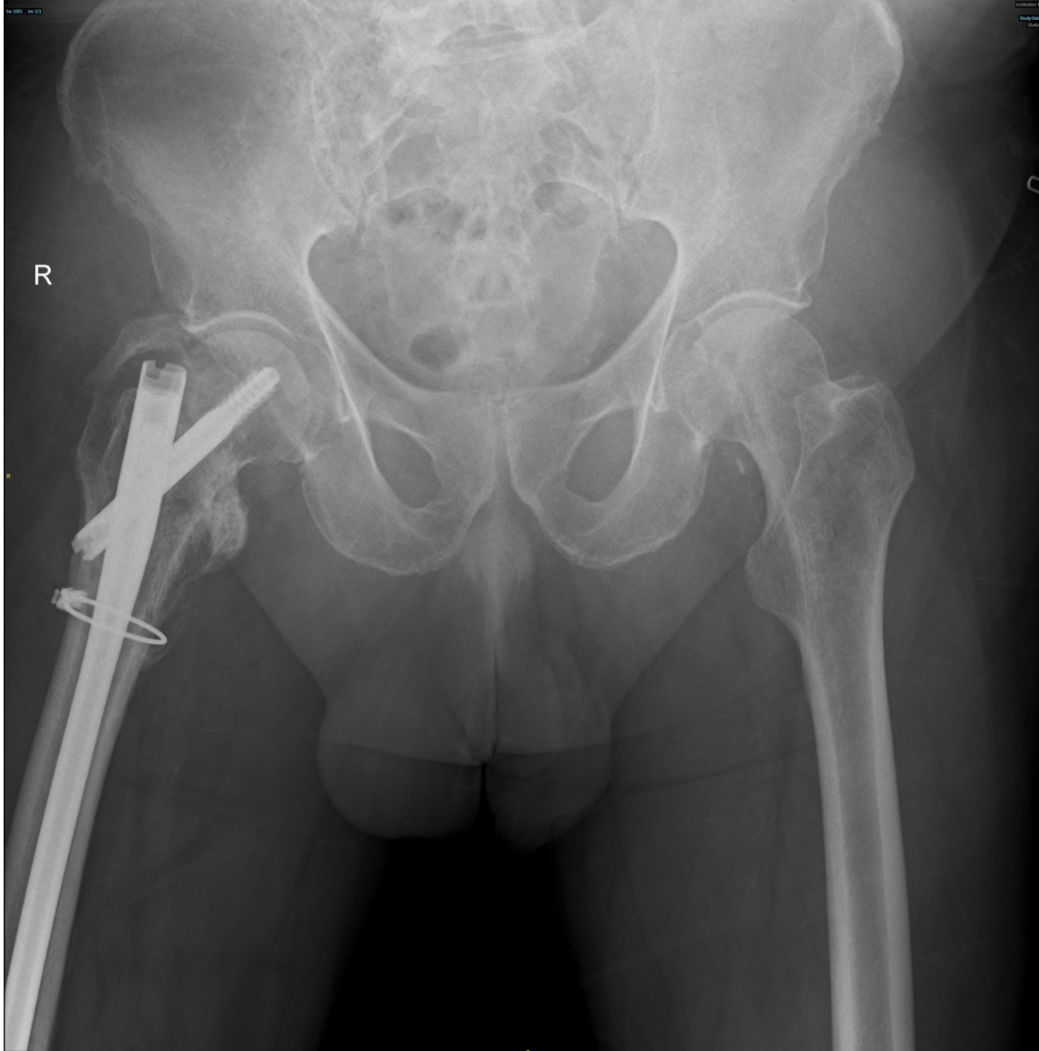
An 83-year-old man was admitted following a mechanical fall at home, having tripped backwards and subsequently developed right hip pain. He normally mobilised with a stick. On examination, the right lower limb was shortened and externally rotated, with neurovascular status intact. X-rays showed a mildly displaced and angulated comminuted intertrochanteric fracture of the right hip, extending into the subtrochanteric region. Both hip joints remained congruent, with early degenerative osteoarthritic changes noted. No other acute bony injury was identified. He underwent operative management with intramedullary (IM) nailing of the right femur using a long gamma nail with cerclage wiring. The immediate post-operative course was initially stable, with blood tests within normal limits.
On day 1 post-operation, he developed acute chest pain associated with significant oxygen desaturation, prompting a medical emergency team (MET) call. Observations at the time showed a NEWS of 9, respiratory rate of 34, oxygen saturations of 78% on room air (requiring 15 L via non-rebreather mask), heart rate of 94 bpm, blood pressure of 118/72, and temperature of 37.7°C. ECG demonstrated new ST segment depression, and serial troponin T levels rose from 236 to 2622. D-dimer was elevated (638), so CT imaging was done which excluded pulmonary embolism and aortic pathology. Blood tests revealed a drop in haemoglobin, prompting transfusion.
Following cardiology review, a diagnosis of Type 2 myocardial infarction (T2MI) was made, likely secondary to anaemia post operatively. He was managed with transfusions, initiation of dual antiplatelet therapy, and intravenous furosemide for associated pulmonary congestion. The patient was transferred to the Acute Care Unit (ACU) for monitoring and stabilisation. Following clinical improvement, he was stepped down back to the orthopaedic ward, where he continued rehabilitation with physiotherapy. Cardiology input advised that no formal outpatient follow-up was required, as the T2MI was attributed to a reversible cause (anaemia), and he was already established on appropriate secondary prevention. Re-referral was advised only if future symptoms suggestive of angina developed.
Discussion:
Patients undergoing hip fracture surgery represent a frail and high-risk perioperative group, and postoperative myocardial injury/infarction is associated with increased mortality [3,4]. In a population-based study, clinically verified myocardial infarction occurred in a notable proportion of patients after hip fracture repair, with most events arising early in the postoperative period [3].
Type 2 myocardial infarction is particularly relevant in the postoperative setting because surgery, blood loss, pain, hypoxia, and systemic stress can all contribute to a mismatch between myocardial oxygen supply and demand [5,6]. In this case, postoperative anaemia was the most likely precipitating factor, with hypoxia and the wider physiological stress response likely contributing further [5,6].
This case highlights the importance of early recognition of clinical deterioration. The presence of chest pain with hypoxia in the immediate post-operative period necessitates urgent escalation, as the differential diagnosis includes myocardial infarction, pulmonary embolism, and pulmonary oedema. Prompt activation of a MET call enabled rapid multidisciplinary assessment and timely investigation.
Management of MI in the post-operative setting presents a significant clinical challenge. Standard therapies, including dual antiplatelet treatment, must be balanced against the increased risk of bleeding following recent orthopaedic surgery. Similarly, fluid management is complex, requiring careful optimisation to treat pulmonary congestion while avoiding hypovolaemia and renal injury.
An additional important consideration is the distinction between Type 1 and Type 2 MI, as this has implications for both management and follow-up. In cases where T2MI is driven by a reversible cause, such as anaemia, treatment is directed at correcting the underlying trigger rather than routine invasive cardiology follow-up.
Overall, this case demonstrates the need for coordinated multidisciplinary care involving orthopaedics, cardiology, and acute medical teams to navigate competing risks and optimise patient outcomes.
Learning points:
- Type 2 myocardial infarction is often precipitated by reversible physiological stressors, particularly anaemia.
- Early activation of a MET call facilitates rapid multidisciplinary input and investigation.
- Multidisciplinary collaboration is essential in guiding treatment and safe recovery.
- Management requires balancing thrombotic risk against post-operative bleeding risk.
References:
- Bhandari M, Swiontkowski M. Management of Acute Hip Fracture. N Engl J Med. 2017;377(21):2053-2062. doi:10.1056/NEJMcp1611090
- Haentjens P, Magaziner J, Colón-Emeric CS, et al. Meta-analysis: excess mortality after hip fracture among older women and men. Ann Intern Med. 2010;152(6):380-390. doi:10.7326/0003-4819-152-6-201003160-00008
- Huddleston JM, Gullerud RE, Smither F, et al. Myocardial infarction after hip fracture repair: a population-based study. J Am Geriatr Soc. 2012;60(11):2020-2026. doi:10.1111/j.1532-5415.2012.04205.x
- Rostagno C, Cartei A, Rubbieri G, et al. Perioperative myocardial infarction/myocardial injury is associated with high hospital mortality in elderly patients undergoing hip fracture surgery. J Clin Med. 2020;9(12):4043. doi:10.3390/jcm9124043
- Thygesen K, Alpert JS, Jaffe AS, et al. Fourth Universal Definition of Myocardial Infarction (2018). J Am Coll Cardiol. 2018;72(18):2231-2264. doi:10.1016/j.jacc.2018.08.1038
- DeFilippis AP, Chapman AR, Mills NL, et al. Assessment and Treatment of Patients With Type 2 Myocardial Infarction and Acute Nonischemic Myocardial Injury. Circulation. 2019;140(20):1661-1678. doi:10.1161/CIRCULATIONAHA.119.040631
