
Introduction
Plica syndrome is an interesting problem that occurs when an otherwise normal structure in the knee becomes a source of knee pain due to injury or overuse. The diagnosis can sometimes be difficult, but if this is the source of your knee pain, it can be easily treated.
This guide will help you better understand
- what a plica is
- how plica syndrome can cause problems
- what doctors can do to treat the condition
Anatomy
What is a plica, and what does it do?
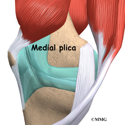
Plica is a term used to describe a fold in the lining of the knee joint. Imagine the inner lining of the knee joint as nothing more than a sleeve of tissue. This sleeve of tissue is made up of synovial tissue, a thin, slippery material that lines all joints. Just as a tailor leaves extra folds of material at the back of sleeves on a shirt to allow for unrestricted motion of the arms, the synovial sleeve of tissue has folds of material that allow movement of the bones of the joint without restriction.
Four plica synovial folds are found in the knee, but only one seems to cause trouble. This structure is called the medial plica. The medial plica attaches to the lower end of the patella (kneecap) and runs sideways to attach to the lower end of the thighbone at the side of the knee joint closest to the other knee. Most of us (50 to 70 percent) have a medial plica, and it doesn't cause any problems.
Related Document: A Patient's Guide to Knee Anatomy
Causes
How does a plica cause problems in the knee?
A plica causes problems when it is irritated. This can occur over a long period of time, such as when the plica is irritated by certain exercises, repetitive motions, or kneeling. Activities that repeatedly bend and straighten the knee, such as running, biking, or use of a stair-climbing machine, can irritate the medial plica and cause plica syndrome.
Injury to the plica can also happen suddenly, such as when the knee is struck in the area around the medial plica. This can occur from a fall or even from hitting the knee on the dashboard during an automobile accident. This injury to the knee can cause the plica, and the synovial tissue around the plica, to swell and become painful. The initial injury may lead to scarring and thickening of the plica tissue later. The thickened, scarred plica fold may be more likely to cause problems later.
Symptoms
What does plica syndrome feel like?
The primary symptom caused by plica syndrome is pain. There may also be a snapping sensation along the inside of the knee as the knee is bent. This is due to the rubbing of the thickened plica over the round edge of the thighbone where it enters the joint. This usually causes the plica to be tender to the touch. In thin people, the tissue that forms the plica may be actually be felt as a tender band underneath the skin. In rare cases where the plica has become severely irritated, the knee may become swollen.
Diagnosis
How will my doctor know it's plica syndrome?
Diagnosis begins with a history and physical exam. The examination is used to try and determine where the pain is located and whether or not the band of tissue can be felt. X-rays will not show the plica. X-rays are mainly useful to determine if other conditions are present when there is not a clear-cut diagnosis.
If there is uncertainty in the diagnosis following the history and physical examination, or if other injuries in addition to the plica syndrome are suspected, magnetic resonance imaging (MRI) may be suggested. The MRI machine uses magnetic waves to show the soft tissues of the body. Usually this test is done to look for injuries, such as tears in the meniscus or ligaments of the knee. This test does not require any needles or special dye and is painless. A computed tomography (CT) scan may also be used to see whether the plica has become thickened. Most cases of plica syndrome will not require special tests such as the MRI or CT scan.
If the history and physical examination strongly suggest that a plica syndrome is present, then arthroscopy may be suggested to confirm the diagnosis and treat the problem at the same time. Arthroscopy is an operation that involves inserting a small fiber-optic TV camera into the knee joint, allowing the surgeon to look at the structures inside the knee joint directly. The arthroscope allows your surgeon to see the condition of the whole knee and determine whether the medial plica is inflamed.
Treatment
What can be done for plica syndrome?
The majority of people with plica syndrome will get better without surgery. The primary goal when treating the plica is to reduce the inflammation. This may require limiting activities like running, biking, or using a stair-climbing machine.
Nonsurgical Treatment
Your doctor may suggest anti-inflammatory medications such as ibuprofen or aspirin to reduce the inflammation. Ice packs or ice massage can help reduce the inflammation and swelling in the area of the plica and may be suggested by your doctor or physical therapist. Ice massage is easy and effective. Simply freeze water in a paper cup. When needed, tear off the top inch, exposing the ice. Rub three to five minutes around the sore area until it feels numb.
A cortisone injection into the plica, or simply into the knee joint, may quickly help to reduce the inflammation around the plica. Cortisone is a powerful anti-inflammatory medication, but it should be used sparingly inside joints. There is always a risk of infection associated with injections into any joint.
Surgery
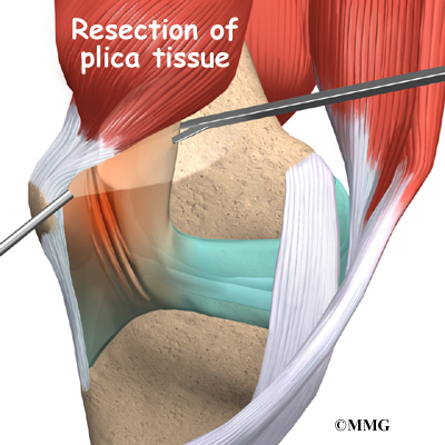
If all nonsurgical attempts to reduce your symptoms fail, surgery may be suggested. Usually, an arthroscope (mentioned earlier) is used to remove the plica. The small TV camera is inserted into the knee joint through one-quarter inch incisions. Once the plica is located with the arthroscope, small instruments are inserted through another one-quarter inch incision to cut away the plica tissue and remove the structure. The area where the plica is removed heals back with scar tissue. There are no known problems associated with not having a plica, so you won't miss it.
Rehabilitation
What should I expect following treatment?
Nonsurgical Rehabilitation
If your treatment is nonsurgical, you should be able to return to normal activity within four to six weeks. You may work with a physical therapist during this time. Treatments involve stretching and strengthening exercises for the leg.
Treatments such as ultrasound, friction massage, and ice may be used to calm inflammation in the plica. Therapy sessions sometimes include iontophoresis, which uses a mild electrical current to push anti-inflammatory medicine to the sore area. This treatment is especially helpful for patients who can't tolerate injections.
After Surgery
Your surgeon may have you work with a physical therapist after surgery. Your first few rehabilitation sessions are designed to ease pain and swelling and help you begin gentle knee motion and thigh tightening exercises. Patients rarely need to use crutches after this kind of surgery.
As the program evolves, more challenging exercises are chosen. Patients do closed chain exercises by keeping their foot on a surface while working the knee joint. These exercises mimic familiar activities like squatting down, lunging forward, and going up or down steps. These exercises help keep pressure off the kneecap while getting a challenging workout for the leg muscles. Your therapist will work with you to make sure you are not having extra pain in your knee during the exercises. You may be shown stretches for the soft tissues along the edge of the kneecap as well as flexibility exercises for the hamstrings, quadriceps, and calf muscles.
The therapist's goal is to help you keep your pain under control, increase the strength of your quadriceps muscles, and maximize the range of motion in your knee. When you are well under way, your regular visits to the therapist's office will end. The therapist will continue to be a resource, but you will be in charge of doing your exercises as part of an ongoing home program.
 |
All content provided by eORTHOPOD® is a registered trademark of Medical Multimedia Group, L.L.C.. Content is the sole property of Medical Multimedia Group, LLC and used herein by permission. |
